Page
The Reproductive System
Completion requirements
View
The role of reproduction is to provide for the continued existence of a species; it is the process by which living organisms duplicate themselves. Animals compete with other individuals in the environment to maintain themselves for a period of time sufficient to enable them to produce tissue nonessential to their own survival, but indispensable to the maintenance of the species.
The Female Reproductive System
The female reproductive system consists of the vulva, vagina, cervix, uterus, fallopian tubes and ovaries.
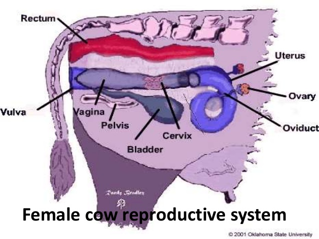
The structure of the female reproductive system
Vulva: The cleft like opening on the female reproductive system is situated just beneath the anus.
Vagina: Continuous with the vulva and lies in the pelvic cavity. The last part of the large intestine (rectum) lies dorsal to it while the urinary bladder lies immediately ventral to it. There is no definite line of demarcation between the vulva and vagina.
Cervix: The cervix is the connection between the vagina and the uterus. The cervix is thick-walled with a narrow tortuous lumen. In the pregnant animal, it is sealed off by a mucous plug.
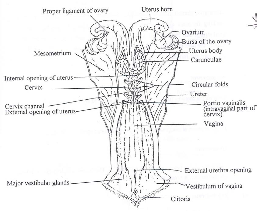
Uterus: The uterus consists of a body of varying lengths between species, and two uterine horns. The horns are attached to the pelvis by the broad ligaments. On the inside of the horns of the uteri of ruminants, the caruncles are found. Normally they are about the size of a pea, but during pregnancy, they enlarge and then have a spongy appearance. These are the sites of attachment between the uterus and the placenta.
Fallopian Tubes: The Fallopian tubes, which transport the fertilised ovum and where fertilisation takes place, are anterior extensions of the uterine horns. At the anterior end of the Fallopian tubes, the ovaries are found. The free end of the tubes is funnel-shaped, the so-called infundibula, which enfolds the ovary at ovulation.
Ovaries: Each female animal has two of these organs, which produce ova. The Graafian follicle containing the ovum is formed in the ovary. When ovulation takes place, the ovum is collected by the infundibulum and moves through the Fallopian tube in the direction of the uterus. Inside the Fallopian tube, the ovum is fertilised by the sperm cells and then moves on into the uterus where it develops into the embryo, later on becoming a foetus.
Oestrus Cycle: The oestrus cycle of the female animal starts at puberty and is associated with distinct physiological changes. These changes, which occur rhythmically during the sexually active season is the only time that the female is fertile.
The Mammary Gland (Udder)
Anatomically the mammary gland is a gland of the skin. In the following discussions, the udder of the cow will be used as an example. The bovine udder consists of four milk glands or quarters. Each quarter is a separate unit with its own duct system, teat cistern and teat. Where the teat canal goes over into the teat cistern there are a series of four to eight radial folds in the mucous membrane. In the mucous membrane of the teat cistern, there are numerous irregular circular and longitudinal folds. The openings of the teats are held closed by a sphincter of smooth muscle and elastic fibres. Basically, the udder of the cow differs from that of other domestic animals as follows:
|
Anatomical Characteristic |
Cow |
Sheep Goat |
Mare |
Sow |
Bitch |
|
Number of functional teats |
4 |
2 |
2 |
10-14 |
8-12 |
|
Number of teat openings per teat |
1 |
1 |
2-3 |
2 |
18-20 |
The following hormones have a direct or indirect influence on the development of the mammary gland:
Thyrotrophic hormone, Growth hormone, ACTH, FSH, LH, and prolactin.
From the posterior pituitary:
- Oxytocin from the hypothalamus via the posterior pituitary plays a very important role in the “let-down” of milk by causing contraction of the smooth muscles and myoepithelial cells of the mammary gland.
- Oestrogen from the Graafian follicle also plays an important role in the development of the mammary gland.
The constituents of the milk are produced directly or indirectly from the blood. Although the osmotic pressure of milk and blood is the same, the constituents of these two fluids differ greatly. Milk contains more sugar, lipids, calcium, phosphorus, and potassium, but fewer proteins, sodium and chloride. The proteins in milk are mainly casein (with small quantities of albumins and globulins), while albumins and globulins are the most important proteins in the blood. The components of colostrum also differ from that of milk. In most animals, colostrum is the medium by which antibodies are transmitted from the mother to the newborn offspring.
Click here to view a video that explains the cow reproductive system.
Last modified: Tuesday, 19 April 2022, 3:19 PM