Page
The Respiratory System
Completion requirements
View
Cavities of the Body
The diaphragm divides the body into two cavities namely:
The thoracic cavity, enclosed by the ribs, contains the heart, lungs, and part of the oesophagus and trachea.
The abdominal cavity contains the stomach, intestines, liver, spleen, pancreas and kidneys. The posterior part of this cavity is called the pelvic area and contains the bladder and the uterus in the female.
Definition:
Diaphragm – A layer of muscle that separates the stomach from the chest and moves up and down when the animal breathes.
The Posterior part – The part pertaining to the rear of the animal.
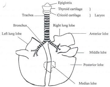
The Respiratory Organs
The respiratory organs supply oxygen and remove carbon dioxide from the blood. The following organs are involved: nasal cavity, turbinate bones, larynx, trachea, lungs (consisting of bronchi, bronchiole and alveoli), the ribs, the thoracic muscles and the diaphragm.
The section from the nose to the bronchi not only serves as airways but also have hair on the mucous membranes, which trap, dust particles, etc. The mucous membrane secretes mucus for taking up particles of dust. The airways also supply heat and moisture to incoming air. Like the arterioles the bronchiole can also constrict or expand thus controlling the inflow of air.
The bronchiole eventually forms alveoli (plural of alveolus). Alveoli are thin-walled structures surrounded by capillary blood vessels. Two very thin membranes thus separate the air in the alveoli from the blood and the exchange of gasses (oxygen and carbon dioxide) takes place through these membranes (see diagram below).
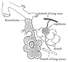
The bronchiole and alveoli of the lungs
The lungs
The lungs are elastic organs, which hang in the thoracic cavity. They are divided into lobes, are spongy and soft, with a pink colour. When an animal is dead, the lung lobe on the lower side of the body will always be darker than the lobe lying above.
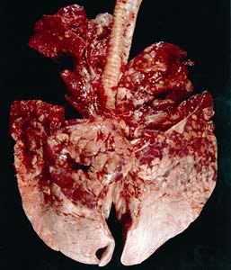
Pneumonia Cow Lung Damaged
Lungs of animals with severe pneumonia as well as the lungs of unborn animals sink when dropped into water. The thoracic cavity is an airtight compartment and when it enlarges a vacuum develops in the thoracic cavity. The lungs are not closed but are in direct contact with the atmosphere through the airways. When the pressure in the thoracic cavity gets less than the atmospheric pressure (due to the enlargement of the cavity) the lungs will fill with as much air as their elasticity will allow. The chest cavity enlarges when the mussels between the ribs, pull the ribs forward. The diaphragm plays an important role in enlarging the thoracic cavity. It consists mainly of muscles and has a domed shaped form which projects into the thoracic cavity. When the muscles of the diaphragm contract the dome flattens and the thoracic cavity enlarges. As soon as the diaphragm and the intercostals muscles relax, the volume of the chest cavity decreases and due to the elasticity of the lungs, air is now expelled from the lungs
Definition:
Mucous membrane – Membrane that secretes a thick slimy liquid that protects the delicate tissues that line certain parts of the body of animals, for example, inside the nose.
Arterioles – A minute arterial branch.
Bronchiole – One of the finer (mm. or less) subdivisions of the branched bronchial tree of the lungs – having no cartilage plate.
Click here to view a video that explains how the lungs work.