Page
The Reproductive System
Completion requirements
View
The Female Reproductive System
The female reproductive system consists of the vulva, vagina, cervix, uterus, fallopian tubes and the ovaries.
Vulva: The cleft like opening on the female reproductive system situated just beneath the anus.
Vagina: Continuous with the vulva and lies in the pelvic cavity. The last part of the large intestine (rectum) lies dorsal to it while the urinary bladder lies immediately ventral to it. There is no definite line of demarcation between the vulva and vagina.
Cervix: The cervix is the connection between the vagina and the uterus. The cervix is thick walled with a narrow tortuous lumen. In the pregnant animal it is sealed off by a mucous plug.
Uterus: The uterus consists of a body of varying length between species, and two uterine horns. The horns are attached to the pelvis by the broad ligaments. On the inside of the horns of the uteri of ruminants, the caruncles are found. Normally they are about the size of a pea, but during pregnancy they enlarge and then have a spongy appearance. These are the sites of attachment between the uterus and the placenta.
Fallopian tubes: The Fallopian tubes, which transport the fertilised ovum and where fertilisation takes place, are anterior extensions of the uterine horns. At the anterior end of the Fallopian tubes the ovaries are found. The free end of the tubes is funnel shaped, the so-called infundibula, which enfolds the ovary at ovulation.
Ovaries: Each female animal has two of these organs, which produce ova. The Graafian follicle containing the ovum is formed in the ovary. When ovulation takes place, the ovum is collected by the infundibulum and moves through the Fallopian tube in the direction of the uterus. Inside the Fallopian tube the ovum is fertilised by the sperm cells and then moves on into the uterus where it develops into the embryo, later on becoming a foetus.
Oestrus cycle: The oestrus cycle of the female animal starts at puberty and is associated with distinct physiological changes. These changes, which occur rhythmically during the sexually active season is the only time that the female is fertile.
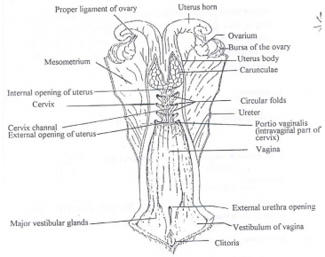
The structure of the female reproductive system
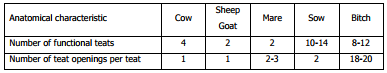
The mammary gland (Udder): Anatomically, the mammary gland is a gland of the skin. In the following discussions the udder of the cow will be used as example. The bovine udder consists of four milk glands or quarters. Each quarter is a separate unit with its own duct system, teat cistern and teat. Where the teat canal goes over into the teat cistern there are a series of four to eight radial folds in the mucous membrane. In the mucous membrane of the teat cistern there are numerous irregular circular and longitudinal folds. The openings of the teats are held closed by a sphincter of smooth muscle and elastic fibres.
Basically, the udder of the cow differs from that of other domestic animals as follows:
The following hormones have a direct or indirect influence on the development of the mammary gland.
- Thyrotrophic hormone, Growth hormone, ACTH, FSH, LH, and prolactin.
- From the posterior pituitary:
- Oxytocin from the hypothalamus via the posterior pituitary plays a very important role in the “let-down” of milk by causing contraction of the smooth muscles and myo-epithelial cells of the mammary gland.
- Oestrogen from the Graafian follicle also plays an important role in the development of the mammary gland.
The constituents of the milk are produced directly or indirectly from the blood. Although the osmotic pressure of milk and blood is the same, the constituents of these two fluids differ greatly. Milk contains more sugar, lipids, calcium, phosphorus, and potassium, but less proteins, sodium and chloride. The proteins in milk are mainly casein (with small quantities of albumins and globulins), while albumins and globulins are the most important proteins in blood.
The components of colostrum also differ from that of milk. In most animals, colostrum is the medium by which antibodies are transmitted from the mother to the new-born offspring.
The Male Reproductive System
The male reproductive system consists of the testis, epididymis, vas deferens, ampullae's, urethra, penis and accessory sex glands.
Testis: The testes (two in normal males) are enclosed in the scrotum. The testis itself is oval shaped and the left and right testes are not always of exactly the same size. Most of the testis is made up of fine tubules in whose walls the spermatozoa are formed. By merging, the tubules become larger and eventually form the epididymis. Spermatozoa undergo their final maturation process in the epididymis and are also stored there.

The location of the parts of the male reproductive system
Epididymis: The epididymis is joined to each testis and consists of a head, body and tail. The head is attached to the dorsal part of the testis. The body, a continuation of the head, goes over into the tail, which lies at the base of the testis.
Vas deferens: The vas deferens are paired ducts that carry semen from the tail of the epididymis, via the spermatic cord, to the ampullae's in the pelvic cavity.
Ampullae's: Basically, these are thickenings of the last part of the vas deferens. Like the vas deferens, the function of the ampullae's is to transport semen, to store semen and to secrete spermatic fluid. They contract forcefully to expel the semen during ejaculation. The ampullae's lie on top of the urinary bladder and open into the urethra.
Urethra: This single duct starts at the bladder and continues right through the length of the penis. In the male it transports both semen and urine.
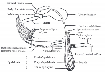
The structure of the male reproductive system
The penis: The male sexual organ used for mating. It lies in an invagination of the skin called the sheath. The penis of a ruminant consists of strong connective and elastic tissue and has a characteristic sigmoid flexure. The penis of the horse is spongier. The tip of the penis is called the glans penis.
Spermatic cord: Consists of the cremaster muscles, blood vessels, nerves and vas deferens. The cavity of the scrotal sac is continuous with the abdominal cavity. A network of closely apposed blood vessels cools down the blood going to the testes. This is necessary because spermatogenesis occurs optimally at temperatures approximately 4 degrees Celsius below body temperature. Apposed Blood vessels packed next to one another to form a cooling network
Accessory sex glands: The functions of these glands are to give volume to the semen and supply nutrients and protection to the spermatozoa. They include the seminal vesicles, prostate and the bulbo-urethral glands.