Page
The Endocrine System
Completion requirements
View
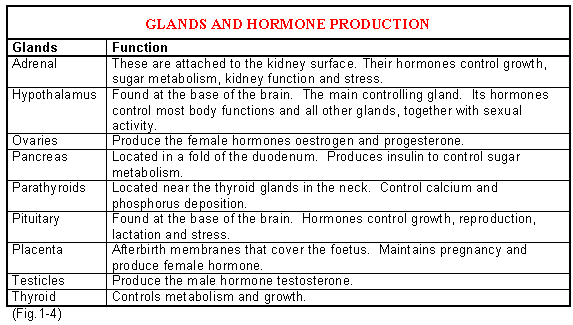
The endocrine system is a system of ductless glands, which secrete chemical substances, called hormones, directly into the blood system. Via the blood circulatory system, they come into contact with the target organ(s) on which they have a specific effect. Hormones have the following general characteristics:
- They regulate reactions rather than initiating them
- They are effective in minimal quantities
- Their levels fluctuate according to the demand
The latter characteristic is necessary for integrated systems, which handle the different requirements of growth, sexual differentiation, reproduction and adaptation to environmental changes.
The Hypothalamus and Pituitary
The pituitary (hypothesis) and hypothalamus are morphologically and functionally intimately associated. They represent the centre of the highest coordination between the endocrine and nervous systems. The hypothalamus is the control centre for the autonomic nervous system whilst the pituitary controls the endocrine system. Together they form a functional unit.
Hypothalamus
The hypothalamus is part of the brain, which lies just below the thalamus, forming the floor of the third ventricle. Numerous releasing hormones or factors are produced by the hypothalamus. These factors are transported to the pituitary where they stimulate the pituitary cells to release their respective hormones into the bloodstream. The hypothalamus has a two-way connection with the cerebral cortex. A close association thus exists between the nervous system (which affects rapid coordination) and the endocrine system, which is involved in the slower chemical coordination of the body.
The Pituitary (Hypothesis) Gland
This small structure lies at the base of the brain and is connected to the hypothalamus by a small pedicle. The part of the pituitary that originates from the brain is called the posterior lobe (neurohypophysis) and the part originating from the upper palate is called the anterior lobe of the adenohypophysis.
Hormones of the Anterior Pituitary
Adreno-corticotropic hormone (ACTH): It is secreted by a stimulus of the corticotrophin-releasing factor (CT- RF) from the hypothalamus. Adreno-corticotropic hormone (ACTH) stimulates the secretion of corticosteroids by the adrenal cortex. On the other hand, corticosteroids inhibit the secretion of ACTH, a process called” negative feedback”.
Growth Hormone (Somatotropin): Secretion is controlled by the growth hormone-releasing factor (GH-RF) from the hypothalamus. The growth hormone controls the general growth of the body (especially the longitudinal growth of the long bones).
Thyrotropic Hormone (TTH): Secretion is controlled by the thyrotropin-releasing hormone (T-Rh). TTH stimulates the growth of the thyroid gland. It controls the uptake of iodine by the thyroid and thus also the synthesis and release of the thyroid hormone.
Follicle Stimulating Hormone (FSH): The releasing hormone from the hypothalamus again stimulates the release of FSH by the anterior pituitary. FSH stimulates the development of the Graafian follicle in the ovary of the female. Oestrogen, which is formed in the follicle, on the other hand, inhibits the secretion of FSH when a certain level is reached.
Interstitial Cell Stimulating Hormone (ICSH): ICSH stimulates the interstitial cells in the testis of the male to secrete testosterone (the male sex hormone).
Luteinizing Hormone (LH): LH-RH causes the release of LH, which is responsible for ovulation in the female animal. Ovulation takes place as soon as a specific balance between oestrogen from the follicle and LH from the anterior pituitary is reached. After ovulation LH also plays a role in the development of the corpus luteum (yellow body).
Prolactin: Prolactin is responsible for the maintenance of secretion of the corpus luteum and is also involved in lactation. In the male, it stimulates the accessory sexual organs.
Hormones of the Posterior Pituitary (Neurohypophysis)
These hormones are formed in the brain and then transported to the posterior pituitary.
Antidiuretic Hormone (ADH): The antidiuretic hormone decreases the volume of urine by increasing water resorption from the filtration solution in the kidney.
Oxytocin: This hormone acts on the mammary gland. It stimulates the flow of milk through its action on the myoepithelial cells and smooth muscle fibres in the mammary gland – the so-called “let-down reflex”. Oxytocin is released as soon as the udder of the cow is stimulated e.g. by the pre-milk washing of the udder. The effect of the hormone only lasts for a few minutes and the cow must therefore be milked as soon as possible, otherwise, there is no proper “let-down” of milk. During parturition (birth) it causes contractions of the uterus muscles and in the act of mating, it aids in the transport of spermatozoa and the ovum. The reaction of oxytocin is inhibited by adrenalin (and thus fear or stress).
The Ductless Glands of the Body
Thyroid
The thyroid consists of two lobes, which are situated on both sides of the trachea, near the larynx. In most domestic animals the lobes are connected. The hormone of the thyroid contains large quantities of iodine, which is the active constituent in this case. The formation of the thyroid hormone is controlled by the thyrotropic hormone (TTH), which is secreted by the pituitary. Thyroid hormone, in turn, inhibits the formation of the thyrotropic hormone, and a balance is thus created between the two hormones in the bloodstream.
With a deficiency of iodine in the body, the formation of thyrotropic hormone is not inhibited, with the result that the continuous production of this hormone causes the thyroid to enlarge. This enlargement is known as goitre. Further symptoms of decreased production of thyroid hormone (or deficiency of iodine) are:
- Obesity – sugars cannot be metabolised fully
- Dwarfism – in young animals
- Hairlessness or loss of hair – in iodine-deficient areas piglets are often born hairless
- Lowered fertility
- Lowered milk production
Parathyroid
The parathyroid consists of two pairs of glands on, or very near, the thyroid glands. They are much smaller than thyroids. Parathyroid hormone controls the uptake of calcium by the intestine and resorption of calcium from the bones, as well as the excretion of phosphorus in the bloodstream. The parathyroid plays a very important role in high calcium intake in dairy cows before the birth of a calf. If the gland’s reaction is lazy, the blood-calcium levels will fall when milk secretion starts, and the cow can get milk fever.
Adrenals
The adrenals lie close to and at the cranial poles of the kidneys. Anatomically a cortex and medulla can be distinguished. Both these areas secrete different hormones.
Cortex: Under the influence of ACTH from the pituitary gland, a series of hormones are formed. The following are the most important:
Mineral corticoid: Controls Na: K balance in the blood and the fluid balance in the body. Loss of minerals through the impaired function of the adrenal cortex causes more water to be excreted by the body with resultant dehydration.
Gluco corticoids: Essential for the control of glucose metabolism (energy) and exert an anti-inflammatory effect.
Medulla:
The hormone adrenalin is produced by the medulla and is responsible for:
- Increased heart rate. More work is done by the cardiac muscles than normal.
- Dilation of the blood vessels of the heart. As a result of the increase in the heart rate and more forceful contractions, a better blood supply is essential.
- Increase in blood pressure. More blood per time unit is forced through the lungs to increase the rate of gas exchange.
- Increased respiration rate.
- Constriction of blood vessels of the muscles.
- Dilation of the blood vessels of the muscles.
- Mucous- and salivary secretion decreases.
- Increase in blood sugar because more energy is used.
All the above prepare the body for action – the so-called “fight-or-flight” reaction.
Pancreas
In addition to the pancreatic enzymes, which are secreted via the pancreatic ducts into the small intestine, this organ also secretes the hormones insulin and glucagon. Impairment in the secretion of insulin causes diabetes mellitus. In this case, an increase in blood sugar is present, the reason being that permeability of cell walls to blood sugar decreases in the absence of insulin, so blood sugar cannot be made available to the tissues. Notwithstanding the fact that kidneys normally reabsorb 100 percent of the glucose in the urine, the blood sugar is so high that the kidneys are not capable of resorbing all the glucose, leading to the excretion of glucose in the urine. Glucagon opposes the action of insulin by raising blood glucose levels.
Permeability: The ability of the cell membrane to transport molecules through.
Ovary: The ovaries are the female reproductive organs that produce ova from the Graafian follicles. A Graafian follicle produces oestrogen. Apart from its role in ovulation (production of ovum) it also causes oestrus.
Testis: The testis is the male organ that produces the hormone testosterone and sperm cells. Testosterone is responsible for the development of secondary male characteristics in the male animal and stimulates the sexual drive.
Thymus: The thymus is involved in the immune system of the body.
Pineal body: The pineal body regulates the sexual activity of seasonal breeders like sheep and horses. The onset of decreasing daylength in autumn stimulates ewes to start cycling. The opposite is true for mares, were increased day length in spring starts the mare cycling.

Click here to view a video that explains the veterinary endocrine system.